Lab News
Lab News
On September 23, 2026 (Wednesday), at 11:00 p.m. Beijing Time (UTC+8), the international journal Cell Metabolism published online the research article "Lactate Couples Metabolic State to Necroptosis Across Pathological and Physiological Conditions" by a team led by Prof. Liming Sun from the Center for Excellence in Molecular Cell Science, Shanghai Institute of Biochemistry and Cell Biology, Chinese Academy of Sciences, in collaboration with Prof. Yingxian Sun from the First Hospital of China Medical University. The study reveals a previously unrecognized mechanism by which the metabolic signal lactate directly couples cellular metabolic state to the necroptotic signaling pathway through RIPK3 lactylation. It further demonstrates that this metabolic cell-death axis plays dual roles in pathological tissue injury and physiological tissue remodeling, providing a new conceptual framework for understanding how metabolic signals govern cell-fate decisions.
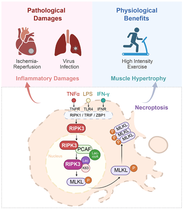
Lactate is a central metabolite generated during glycolysis. In recent years, it has emerged as an important metabolic signaling molecule whose biological functions have been studied primarily in the context of cancer. However, how lactate translates metabolic state into cell-fate decisions—particularly how it directly regulates necroptosis under physiological and non-cancer pathological conditions—has remained largely unknown.
In this study, the researchers first performed a small-molecule library screen and identified lactate as a key regulator of necroptosis. Mechanistically, lactate promotes lactylation of RIPK3 at lysine 63 (K63), thereby directly coupling cellular metabolic state to the necroptotic signaling pathway. The acetyltransferase PCAF catalyzes RIPK3-K63 lactylation, which enhances RIPK3 kinase activity and promotes its interaction with the downstream executioner MLKL, ultimately triggering necroptosis. In contrast, substitution of K63 with the non-lactylatable mutant (RIPK3-K63R) markedly reduced RIPK3 kinase activity and significantly suppressed necroptosis, identifying RIPK3-K63 lactylation as a critical molecular switch controlling necroptotic signaling.
To determine whether inhibition of RIPK3-K63 lactylation could protect tissues from pathological injury, the researchers employed mouse models of myocardial ischemia-reperfusion injury and influenza virus infection. RIPK3-K63R knock-in mice exhibited significantly reduced myocardial infarct size following ischemia-reperfusion injury. Pharmacological inhibition of PCAF attenuated cardiac injury by suppressing RIPK3 lactylation. In the influenza infection model, RIPK3-K63R mutant mice displayed markedly reduced lung pathology, slower body-weight loss, and significantly improved survival. Together, these findings demonstrate that RIPK3-K63-lactylation-driven necroptosis is a major driver of tissue injury in ischemic and viral diseases, highlighting this modification as a promising therapeutic target for organ protection.
Remarkably, the same metabolic cell-death mechanism performs a completely different physiological function. During high-intensity exercise, the large amount of lactate generated in skeletal muscle induces RIPK3-K63 lactylation and moderately activates necroptosis in myofibers. This controlled necroptotic response promotes activation and proliferation of muscle stem cells (MuSCs), thereby facilitating post-exercise skeletal muscle regeneration and remodeling.
Overall, the study identifies lactate as a metabolic signaling molecule that directly regulates necroptosis through RIPK3 lactylation, establishing a previously unrecognized "metabolite–post-translational modification–necroptosis" regulatory paradigm. By demonstrating that this metabolic cell-death axis drives tissue injury under pathological conditions while promoting tissue remodeling under physiological conditions, the work provides a new conceptual framework for understanding how metabolic state governs cell-fate decisions. These findings not only advance our understanding of the metabolic regulation of regulated cell death but also offer new opportunities for therapeutic intervention in ischemia-reperfusion injury, viral infectious diseases, and regenerative medicine.
The study was co-corresponded by Prof. Liming Sun from the Center for Excellence in Molecular Cell Science, Chinese Academy of Sciences, and Prof. Yingxian Sun from the First Hospital of China Medical University. Prof. Naijin Zhang, Dr. Xinyue Huang, Dr. Lucerne Lu, Dr. Yuanming Zou, and Dr. Guozhen Sun are the co-first authors. This work was supported by the National Natural Science Foundation of China, the Ministry of Science and Technology of China, the Chinese Academy of Sciences, the Department of Science and Technology of Liaoning Province, and the core facilities of the Center for Excellence in Molecular Cell Science.
Reference: doi.org/10.1016/,.cmet.2026.08.022
